January 4, 2025 Graphene, Scientific alternative studies
The very purpose of the scientific method is to make sure that nature has not led you to believe that you know something that you do not know.
Graphene Oxide, Neuroinflammation and Neurodegenerative Diseases
Reference Study
Chen, HT; Wu, HY; Shih, CH; Jan, TR (2015). A differential effect of graphene oxide on the production of proinflammatory cytokines by murine microglia. Taiwan Veterinary Journal, 41 (03), pp. 205-211. https://doi.org/10.1142/S1682648515500110
Facts analyzed
Microglial cells are specialized neuroimmune cells found in nerve tissue.
Their function is similar to that of phagocytes, so they are responsible for removing substances and waste, including tumors, microorganisms and invasive agents.
When activated by any damage to the brain or nervous system, they secrete so-called cytokines, as explained for example by (Albarzanji, Z. N.; Mahmood, T. A.; Sarhat, E. R.; Abass, K. S., 2020) and (Rizzo, P.; Dalla-Sega, F. V.; Fortini, F.; Marracino, L.; Rapezzi, C.; Ferrari, R., 2020).
The study showed that a murine microglia (a rodent similar to a mouse) treated with reduced graphene oxide at a dose of (1-25 μg/ml) for 24 h produced pro-inflammatory cytokines and suppressed the production of -1β (i.e. interleukin).
Interleukin is a cytokine whose function within the immune system is to regulate activation, proliferation, and antibody production, as well as to mark where they need to go to do their job.
In other words, graphene oxide interferes with the normal functioning of the immune system, causing it to be inhibited or dysfunctional.
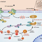
In addition, the study states that “lysosomal permeability and alkalinity are increased in GO-treated microglia, while cathepsin B and ICE activity are decreased.
Taken together, these results demonstrate that GO exposure differentially affected the production of pro-inflammatory cytokines associated with modulation of the lysosomal cytokine processing pathway“.
This statement provides important details.
The first is the increase in microglial alkalinity.
This is by no means trivial, since an increase in alkalinity at the level of brain cells or the nervous system necessarily leads to a low pH that can cause psychiatric and neurodegenerative disorders, as pointed out in the John Hopkins University study (Prasad, H.; Rao, R., 2018), which is directly related to the previous analysis on graphene oxide and its ability to cross the blood-brain barrier.
Second, the activity of cathepsin B (a protein responsible for breaking down the amyloid plaque proteins responsible for Alzheimer’s symptoms) and ICE (the interleukin-converting enzyme IL-1β) was reduced, affecting their proper functioning.
Third, it also affected the modulation of the lysosomal pathway, the lysosomal degradation process that affects proper cellular function.
A review of the scientific literature revealed recent evidence that neuroinflammation caused by “activated microglia and astrocytes may contribute to the progression of pathogenic damage to neurons in the substantia nigra (SN)“.
Similarly, oxidative stress “can be caused by various stressors, such as environmental pollutants or mitochondrial dysfunction“, see (Dowaidar, M. 2021).
This statement fits with the observations in the article discussed in this post, that graphene oxide causes microglia activation.
It also fits with the work of (Prasad and Rao, 2018), which affects astrocyte ApoE4 acidification (astrocytes are glial cells responsible for the development of the central nervous system, among other functions).
The same is true for the work of (Alpert et al., 2020), who analyze clinical cases of depression related to cytokines or cytokine storm.
Conclusion
The article shows that “GO” graphene oxide causes changes in the microglial cells of the central nervous system that affect the functioning of the immune system.
This greatly reduces the ability to cope with infection and disease, leaving the animal or person inoculated with graphene oxide in a vulnerable situation to any biological or chemical contingency or risk.
In addition to alterations in the immune system, neurological damage, oxidative stress and mitochondrial dysfunction (due to their inability to maintain homeostasis) are noted, as well as a reduction in interleukin and ICE levels, which in turn cause alkalinity in microglia, which can lead to neurodegenerative diseases.
Thus, it can be concluded that the potential presence of graphene oxide in so-called “vaccines” can induce neuroinflammation, promote the development of neurodegenerative diseases due to alkalinity and low pH in brain tissue, and cause permanent neurological damage.
Bibliografia
1.Albarzanji, ZN; Mahmood, TA; Sarhat, ER; Abass, KS (2020). Cytokines Storm Of COVID-19 And Multi Systemic Organ Failure : A Review. Systematic Reviews in Pharmacy,, 11 (10), pp. 1252-1256.
2.Alperto, O.; Iniziato, L.; Garren, P.; Solkhah, R. (2020). Cytokine storm induced new onset depression in patients with COVID-19. A new look into the association between depression and cytokines-two case reports. Brain, Behavior, & Immunity-Health, 9, 100173. https://doi.org/10.1016/j.bbih.2020.100173
3.Dowaidar, M. (2021). Neuroinflammation caused by activated microglia and astrocytes can contribute to theprogression of pathogenic damage to substantia nigra neurons, playing a role inParkinson’s disease progression. https://osf.io/preprints/ac896/
4.Prasad, H.; Rao, R. (2018). Amyloid clearance defect in ApoE4 astrocytes is reversed by epigenetic correction of endosomal pH. Proceedings of the National Academy of Sciences, 115 (28), pp. E6640-E6649. https://doi.org/10.1073/pnas.1801612115
5.Rizzo, P.; Dalla-Sega, FV; Fortini, F.; Marracino, L.; Rapezzi, C.; Ferrari, R. (2020). COVID-19 in the heart and the lungs: could we Notch the inflammatory storm ? Basic Research in Cardiology, 115 (31). https://doi.org/10.1007/s00395-020-0791-5

